published · edmund optics case study
edmund optics published our case study, a low-cost, handheld retinal camera for screening premature infants in low-resource settings, in their knowledge center. the four of us on team retinex wrote it: the illumination, optics, capture and control subsystems, the USAF 1951 resolution results, and where the project goes next.
edmund optics also donated the glass fiber-optic light guide the device uses, which keeps the LED's heat away from the infant's face and makes coaxial illumination practical at this cost.
the problem
retinopathy of prematurity (ROP) is a condition where abnormal blood vessels grow in the retinas of babies born before 36 weeks or under 1,500g. left untreated, those vessels contract and pull the retina off the back of the eye. permanent blindness.
here's the thing: if you catch it early, laser therapy can prevent vision loss entirely. the problem is that the screening cameras that do this cost $14k–$125k, require a trained ophthalmologist to operate, and are too bulky to move between hospitals. in countries like kenya, where neonatal survival rates have jumped but specialist infrastructure hasn't kept up, countless infants go undiagnosed.
the scale of it: about 15 million babies are born prematurely every year. a hospital study in kenya found 41.7% of screened premature infants had ROP, a rate comparable to high-income countries, in a place where the screening infrastructure barely exists. kenya has roughly 115 ophthalmologists for a population of 55 million, more than half of them in nairobi. the standard wide-field camera, the RetCam, runs upward of $125,000, and the hospitals that have one often share it between institutions. the problem isn't awareness. it's access.
design an affordable, portable retinal screening device that a neonatal nurse with one hour of training can use. so hospitals in low-resource settings can finally screen for ROP before it's too late.
by the numbers
the camera is already sufficient for detecting plus disease. the vessel dilation and tortuosity in zone I that signals ROP. zone I is where the disease is most dangerous, and our static image captures it cleanly. future iterations will use image stitching to push the FOV past 120° (the full ISO target for ROP screening).
the device
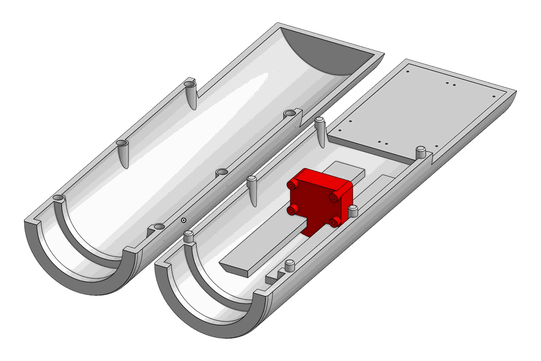
the final prototype is a handheld 3D-printed tube built around four subsystems: illumination (a high-output warm LED), magnification (a 28D ophthalmic lens), capture (a 64MP arducam owlsight camera with motorized autofocus and adjustable exposure), and control (a raspberry pi zero 2W). the form factor stays compact and the component count low, which is what keeps it cheap and keeps it usable one-handed at a bedside.
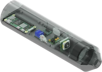
the CAD with the housing ghosted: pi at the back, LED driver and fiber coupling mid-tube, camera on its autofocus mount, 28D lens in the front cone
how the light gets in
illuminating a premature infant's retina is a set of competing constraints. you need enough light to image through a tiny pupil and resolve fine vessels at the back of the eye. a heat-generating light source can't sit near the infant's face. and specular reflections off the cornea and lens will wash out the very structures you're trying to see.
so the LED lives at the back of the device and a glass fiber-optic light guide, donated by edmund optics, carries the light to the imaging head. that physically separates the heat from the front of the device, and at the head the light enters the optical path coaxially, aligned with the imaging axis, which largely eliminates the corneal and lens reflections. the fiber guide is what makes the coaxial layout practical at this cost: enough optical throughput to light the pupil efficiently, in a package that stays compact. PWM brightness control is being integrated, and a hands-free foot pedal for use during imaging is in development.
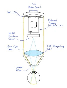
the optical path: LED, fiber guide, coaxial illumination, 28D lens, retina, sensor
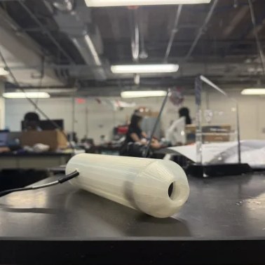
the final prototype with the fiber-optic illumination integrated
the 28D lens is the same type used in standard indirect ophthalmoscopy, chosen because it gives a field of view over 60° inside the device's compact geometry. the 64MP sensor matters because the early signs of ROP, vessel dilation, tortuosity and ridge formation, are fine detail that lower-resolution sensors miss at this magnification.
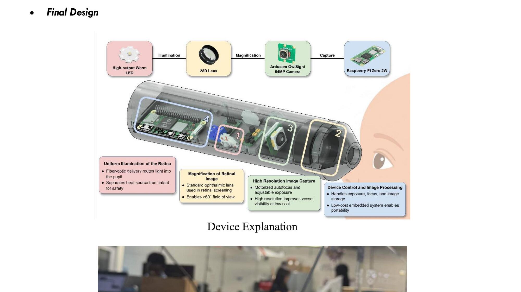
the four components, from pupil to processor
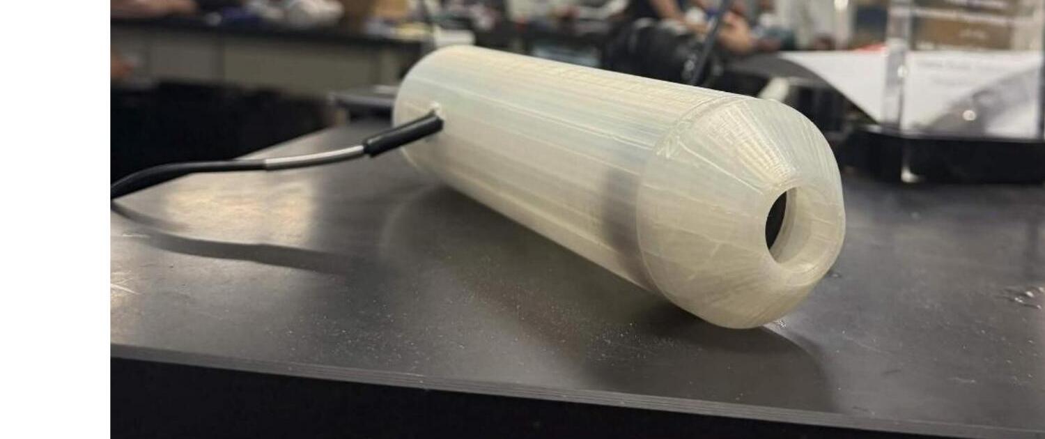
the assembled device: single-piece 3d printed housing, swappable front cone
the app
the raspberry pi manages every subsystem: camera exposure and focus, LED brightness, and the USB link to the companion app. the app gives a live feed for positioning, one-tap capture, a structured image gallery and patient records, in english and kiswahili, so a nurse can capture, label and review screenings without ever leaving the patient. the target is a nurse with no imaging experience, operable after a short orientation.
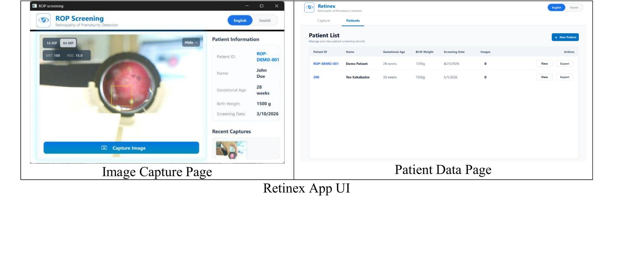
image capture (left) and patient management (right)
the journey
we inherited a prototype called IRIS from a previous EDES 120 team. it was a great starting point but had a few critical issues: bulky casing, external lighting causing glare, blunt front end that made pupil alignment hard, low camera resolution. so we redesigned around those.
inherited prototype analysis
tested the IRIS prototype and ran its AI model. measured baseline resolution at 8.0 lp/mm. far below clinical needs. identified key failure points.
medium-fidelity prototype
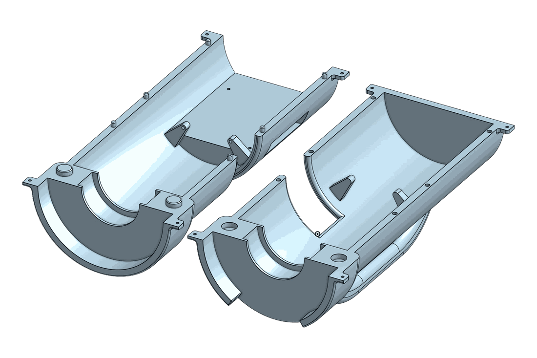
my main contribution: designed and CADed a smaller, ergonomic housing with autofocus and a higher-resolution camera. measured 32 lp/mm, a 4× jump from IRIS.
final prototype
refined optics, moved the warm LED to the back of the tube with a fiber-optic light guide feeding the imaging head, tuned autofocus, integrated the raspberry pi. final resolution: 35.9 lp/mm, FOV 60–80°, plus disease detection working.
handoff to client
delivered working device + documentation to our client katherine sammy, and handed off all materials for the next team.
where we started: the inherited IRIS prototype
mid-cycle redesign: smaller, ergonomic, autofocus
testing
i designed and CADed a custom testing rig that holds the prototype, slides the camera for focus testing, and aligns it precisely with a USAF 1951 resolution target the standard for measuring optical resolution.
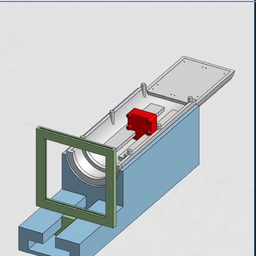
testing rig: gray = prototype holder, red = camera slider, blue = base, green = test plate holder
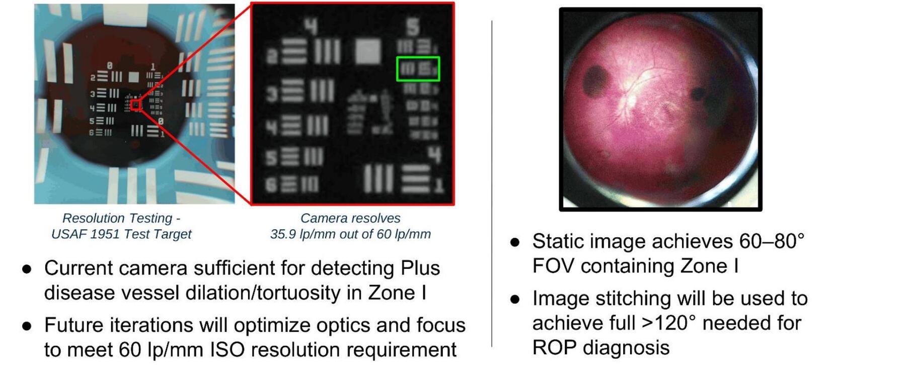
final resolution test (35.9 lp/mm) and a captured retina image with 60–80° FOV containing zone I
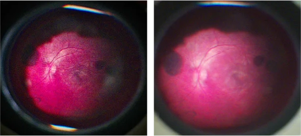
early imaging tests on the model retina: the optic disc and vessels resolve cleanly at the centre of the field
the ISO 15004-2 standard for ophthalmic imaging instruments asks for 60 lp/mm. at 35.9 the prototype is already expected to resolve the vessel dilation and tortuosity that define plus disease in zone I, the innermost region and the site of the most severe ROP. reaching that in a first-semester prototype built for under $200 is the foundation the next iterations build on.
what i worked on
i focused on the hands-on engineering side: the physical device and the test infrastructure.
- cad + mechanical design. designed every iteration of the housing, internal mounts, ergonomic form factor, and the swappable front cones.
- prototyping. built and assembled the physical prototypes: fit, tolerance, integration of the camera, lens and pi inside the casing.
- testing rig design. the entire rig: prototype holder, adjustable camera slider, base and integrated x-axis test plate holder.
- optical testing. ran the USAF 1951 resolution tests, documented results, iterated on the optics.
- optics research. contributed to lens selection and modeling the premature infant eye geometry.
bill of materials
under $500 was our target. we landed at $154.64.
| component | cost |
|---|---|
| arducam owlsight 64MP camera | $55.99 |
| pocketretina 28D BIO lens | $49.99 |
| raspberry pi zero 2W | $15.00 |
| 3W high-output LED | $10.19 |
| buck-boost converter | $9.99 |
| fiber optic cables | $7.99 |
| high-power MOSFET trigger switch | $5.49 |
| total | $154.64 |
what's still hard
we shipped a working prototype but we're not pretending it's done. these are the real challenges the next team will pick up:
- no real-world clinical validation yet. we tested on a model retina. dilated infant eyes will behave differently: movement, pupil response, the works. clinical validation through IRB approval is the most important next step.
- the illumination still needs validation. the fiber-optic coaxial arrangement works, but light delivery efficiency, illumination uniformity and ISO 15004-2 retinal photosafety under real exposure conditions haven't been formally verified. alternative light-guide configurations and coupling methods are on the list.
- focus distance bottleneck. the camera has a 12 cm minimum focus distance, which means ~15 cm of wasted space inside the tube. swapping in a different sensor with a shorter minimum could reclaim that.
- autofocus tuning. the autofocus settle time and fine clarity still need adjustment for the screening workflow.
- FOV to 120°. the static image hits 60–80°. getting to the full 120° needed for ROP diagnosis means building an image stitching pipeline.
the good news
we came in at $155 against a $500 budget. that leaves a lot of headroom for the next team to put real money into the lens or a better sensor without breaking the affordability goal.
recommendations for the next team
- prioritize clinical validation. get IRB approval and capture images on real dilated infant eyes, that's the single biggest unknown.
- run formal user studies. we got informal feedback from judges and an ophthalmologist; the next team should run structured surveys with target users before further design changes.
- validate the illumination. characterise the edmund optics light guide's uniformity and throughput, try alternative coupling methods, and verify photosafety compliance before anything goes near a real patient.
- hardware swap for the focus distance. look at sensors with shorter minimum focus distances to reclaim the wasted internal length.
- spend some of the budget headroom. $345 of room: a higher quality lens (~$500) might be worth it; partnering with thorlabs or edmund optics could bring that down.
where it goes next
the project continues through future rice design teams. the roadmap we published with edmund optics, in order:
- image quality. resolution toward the 60 lp/mm ISO benchmark, coverage beyond 120° through multi-image stitching and better optical alignment. all achievable inside the current architecture.
- illumination. uniformity, coupling, and ISO 15004-2 photosafety compliance under clinically relevant exposures.
- clinical validation. an IRB-approved study, hospital partnerships, and comparative imaging against existing screening systems.
- usability. finishing the hands-free foot pedal, shortening the imaging procedure, and folding in structured feedback from clinicians in kenya, especially for one-handed bedside operation.
- the long game. low-cost portable imaging plus telemedicine workflows, and eventually AI-assisted triage of high-risk cases.
handoff
we already delivered a demonstration to our client, katherine sammy, walked her through the device, and showed her the poster. all four of us would love to help the next team that picks this up.
team retinex
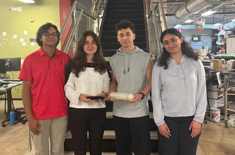
team retinex at the OEDK with the prototype. two freshmen, two sophomores, one semester
samer marmash, alisha sankhe, teo kakabadze, clin vadakkal shaiju. EDES 200 engineering design, Rice University. the four of us also wrote the edmund optics case study ↗.
client: katherine sammy · sponsor: harrell huff · mentors: dr. oden, dr. hunter, dr. ghosn, dr. emmanuel chang. support: oshman engineering design kitchen + donations from thorlabs and edmund optics.
← back to projects